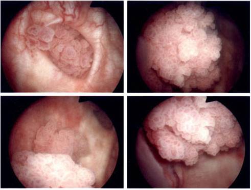
The bladder is an organ located in the pelvic cavity that stores and discharges urine. Urine is produced by the kidneys, carried to the bladder by the ureters, and discharged from the bladder through the urethra. Bladder cancer accounts for approximately 90% of cancers of the urinary tract (renal pelvis, ureters, bladder, urethra).


Endoscopic view of bladder cancer
BLADDER CANCER
FREQUENTLY ASKED QUESTIONS
What causes bladder cancer?
Over 90 percent of people who develop cancer of the urinary bladder have smoked cigarettes at some point in their lives. In addition, bladder cancer is three times more likely to occur in men, further evidence of its association with cigarette smoking. Even though one may have stopped smoking for 20 or 30 years, the effects of tobacco, having been concentrated in the urine over time, continue to adversely impact the urinary system.
Another cause of bladder cancer is radiation to the lower abdomen. Thus, men who have had radiation therapy for prostate cancer may develop bladder cancer many years later. Individuals exposed to industrial dyes and/or chemicals may also have a higher incidence of bladder cancer.
How can you determine whether the cancer has spread?
Bladder cancer is heterogeneous in nature. Many bladder tumors are “low grade” and are not likely to penetrate the wall of the bladder. Because these tumors do not spread, some have argued that they are not really cancerous. These papillary tumors, confined to the surface of the bladder, often recur following removal. But they very infrequently invade the wall of the bladder and, again, are not life threatening.
Other tumors are “high grade” and may grow deep into the wall of bladder and spread to other organs. It is possible for us to learn about the extent of the cancer by removing the tumor and examining it to determine if the cancer has invaded the bladder wall. Once “high grade” tumors have demonstrated the ability to invade the wall, they can potentially spread to other parts of the body.
What is a cystoscopy?
A cystoscopy allows the urologist the opportunity to look into the bladder with a lighted instrument to see if there are any tumors. A local anesthetic is used to minimize the discomfort and one dose of an antibiotic is given following the procedure.
Why do tumors of the bladder tend to occur again following initial removal?
Despite removal of the initial tumor, there is at least a 50 percent likelihood a patient will develop another tumor. If this happens, it usually does so within the first year. There are three reasons why tumors can recur: 1) the carcinogen (e.g., cigarette smoking) has affected the entire bladder surface and, given sufficient time, initiates the growth of another tumor; 2) implantation or “seeding”, following “transurethral resection” (removal of the initial tumor by endoscopic means) may allow remaining microscopic tumor cells, floating within the bladder, to land on a fertile surface, and develop into the next tumor; and 3) incomplete removal of the initial tumor(s).
Regular monitoring of the bladder is necessary to determine whether a new tumor has developed. This monitoring consists of cystoscopy (looking into the bladder with a cystoscope) and cytology (examining the urine for the presence of tumor cells.
Are there any ways to prevent new tumors from forming?
Anti-cancer medicines, placed into the bladder (intravesical therapy) can be an effective way to reduce the likelihood of a new tumor following endoscopic removal of the initial tumor. Intravesical therapy may be either chemotherapy or immunotherapy. The most frequently used chemotherapeutic drug is Mitomycin C and is instilled into the bladder immediately after tumor removal.
The most common method of immunotherapy is placing the drug “BCG” into the bladder. The patient must wait for two weeks following the surgical removal of the tumor before beginning BCG, which is instilled weekly for six consecutive weeks.
Most importantly, if you have not stopped smoking, do so at once. If anyone around you smokes cigarettes, ask them to stop.
Are there methods to determine if there is a bladder tumor without looking into the bladder with a cystoscope?
At the present time, periodic cystoscopy is the standard of care. One may supplement the endoscopic examination with cytology (looking for tumor cells in the urine) or with a tumor marker. Cytology is likely to identify high-grade tumors but may not recognize low-grade tumor cells. Tumor markers are substances that are preferentially found in the urine as a result of the presence of tumor cells, but, unfortunately, are not specific for bladder cancer. Consequently, cystoscopy is a necessary monitoring tool.
What are the options if a tumor invades the wall of the urinary bladder?
Once a tumor has demonstrated the ability to invade the muscle of the bladder, local resection by a Transurethral Resection (TUR) is no longer successful in removing the entire tumor. Two remaining approaches are 1) removal of the urinary bladder or 2) a combination chemotherapy and radiation.
Removal of the bladder, or cystectomy, includes removal of the prostate in the man and the uterus in the woman. The reason to remove these is that the cancer may grow into these adjacent organs and, because of the usual age of the bladder cancer patient, may be more important for survival and less critical to quality of life.
If the urinary bladder is removed, how does the urine get out of the body?
There are two primary alternatives for urinary diversion. If one elects to have an ileal conduit, a portion of the small bowel (ileum) is detached and one end is connected to the skin at the abdomen as a stoma while the two tubes from the kidney (the ureters) are attached to the other end. A small appliance is attached at the side of the abdomen to collect the urine.
The other possibility is to have a relatively complicated reconstruction called a neobladder. 40 to 50 cm (approximately 20 inches) of ileum are separated from the rest of the bowel and reconfigured in the shape of a bladder. One portion of this “neobladder” is attached to the urethra, the normal passageway from the bladder for urine, and the ureters are attached at the other end. Once this is constructed, it is possible for the patient to urinate resembling his/her normal voiding pattern. Unfortunately, the neobladder does not have the same muscle wall as the normal bladder and 15 percent of patients will have to catheterize themselves every six to eight hours to empty their urine.
Is this procedure to remove the urinary bladder a major operation and what will determine which urinary diversion is most appropriate for me?
First of all, a cystectomy, removal of the bladder, is a major operation. One to five percent of patients may die from the surgery. Age and a number of other factors increase the risk of the surgery. The operation itself normally takes four to six hours. Patients who have medical problems such as heart or lung disease or who are overweight are at greater risk. It is also critical that the bowel tract work properly after the procedure, as an obstruction or blockage of the bowel is one of the most frequent postoperative complications.
When is intravenous chemotherapy used for bladder cancer?
Chemotherapy is used when the cancer is known to have spread to other parts of the body (metastasis) or is likely to have spread. In this case, chemotherapy may be given prior to surgery to enhance a patients’ prognosis. But chemotherapy, in combination with radiation, is also used as a bladder preservation strategy.
If I elect the bladder preservation strategy, does that prevent me from ever having my bladder removed?
Chemotherapy alone does not preclude bladder removal surgery. When one adds radiation therapy to the equation, the surgery becomes much more difficult, enhancing the risk of complications. Therefore, the patient most commonly chooses between cystectomy and the bladder preservation strategy.
Can this type of cancer also affect the kidneys?
Because the lining of the kidneys and ureters are the same as that of the bladder, the cancer-causing agent can also affect these structures. Consequently, the kidneys must be monitored by performing an IVP (kidney x-ray) to make certain that no tumor has developed. If the tumors in the bladder are low-grade, this risk of a tumor in the kidney is small; in tumors that are high-grade, the risk increases.
STRENGTHENING YOUR PELVIC FLOOR MUSCLES
![]() Click here to download the Bladder Health Control Information
Click here to download the Bladder Health Control Information